The right scan depends on your gestational age, symptoms, pregnancy history and the clinical question your obstetrician needs answered. By the end, you will be able to match common scans to their timing, understand the measurements and preparation involved, and compare Parel clinics without choosing a package blindly.
Key takeaways
- Use a dating scan when your dates are uncertain, cycles irregular, or pain or bleeding occurs.
- Choose screening tests based on what they detect, not the scan package name.
- Book anatomy, growth or Doppler scans only when timing and clinical purpose match.
- Compare Mumbai clinics by qualified staff, reporting and follow-up—not bundled scans.
Start with gestational age: which scan is needed first?
Choose a dating and viability scan when your last menstrual period is unknown or unreliable, your cycles are irregular, conception followed IVF, or you have pain or bleeding. Before about 10 weeks, a transvaginal ultrasound scan for pregnancy often shows the uterus and embryo more clearly than an abdominal scan.
It can locate the pregnancy, assess viability, measure crown-rump length for dating and investigate ectopic pregnancy; it is not automatically needed for every uncomplicated pregnancy.
ScanTimingMain questionDating and viabilityUsually before 10 weeksWhere is the pregnancy, is it developing, and how far along is it?Nuchal translucency11+0 to 13+6 weeks, with crown-rump length 45–84 mmHow much fluid is behind the fetal neck for first-trimester screening?Early anatomySelected cases before the mid-trimester scanAre selected structures visible? It cannot replace the detailed anatomy examination.
If a reliable first-trimester date exists, do not routinely reset the due date from later scans. Fetal size naturally varies, and growth restriction can make a fetus measure smaller.
IVF dating, twins, previous miscarriage, bleeding or pain can change scan timing and frequency. Ask your obstetrician to name the clinical question rather than booking a generic package. If you are searching “which obstetric ultrasound scan is right for me in mumbai,” start with gestational age and symptoms, not a 3D or 4D label.
Match screening tests to what they can actually detect
Choose a routine ultrasound scan for pregnancy, nuchal translucency, combined first-trimester screening or NIPT according to the question you need answered, not the package name.
OptionWhat it detectsWhat it cannot confirmRoutine ultrasoundPregnancy location, anatomy, growth and fetal featuresEvery chromosomal condition or normal future developmentNuchal translucencyFluid behind the fetal neck at 11+0 to 13+6 weeks, with crown–rump length 45–84 mmA diagnosis of a chromosome conditionCombined screeningNuchal translucency, maternal age, pregnancy-associated plasma protein-A and free beta-hCG to estimate riskCertainty; a high-risk result needs further assessmentNIPTCell-free DNA in maternal blood to screen for selected chromosomal conditionsStructural abnormalities or a definitive diagnosis
Choose the screening path with your obstetrician. A normal NIPT does not rule out heart, brain, spine or other structural abnormalities, while a normal ultrasound does not rule out every chromosomal condition.
The detailed anomaly, or level-II, scan is usually performed at 18–22 weeks. It examines the brain, spine, face, heart, abdomen, kidneys, limbs, placenta and amniotic fluid. It remains a screening examination, not a guarantee.
- Ask for diagnostic testing or targeted imaging with a fetal-medicine specialist if screening or ultrasound raises concern.
- Keep the anomaly scan even after a low-risk NIPT; the tests answer different questions.
- Do not replace a medically indicated two-dimensional examination with a 4D or HD keepsake package. Extra visual effects do not make the assessment more complete.
Know when anatomy, growth and Doppler scans are appropriate
The anomaly scan establishes the mid-pregnancy structural assessment; later imaging answers a specific concern rather than repeating it automatically. A growth scan measures biparietal diameter, head circumference, abdominal circumference and femur length, then estimates fetal weight. It also checks growth trajectory, presentation, placenta and amniotic fluid.
Estimated fetal weight has an error margin, so one number is not your baby’s exact weight.
ScanClinical questionWhat it assessesGrowth scanIs growth or delivery planning a concern?Biometry, estimated weight, trajectory, presentation, placenta and fluidUmbilical-artery DopplerIs placental blood flow affected?Blood-flow indices when fetal-growth restriction, hypertension, diabetes, abnormal placental function or another defined concern existsCervical-length assessmentIs there preterm-birth risk or a short cervix?Cervical length, often by transvaginal ultrasoundBiophysical profileHave movements reduced?Movements, tone, breathing and fluid, combined with fetal-heart-rate monitoringPlacental follow-upIs the placenta low-lying or covering the cervix?The placental edge’s distance from the cervix; transvaginal measurement is more accurate
Umbilical-artery Doppler is not a universal add-on for a normally growing, uncomplicated pregnancy. Near delivery, an ultrasound scan for pregnancy may check presentation, placenta, fluid and growth when those findings affect planning. Twins need first-trimester assessment of chorionicity and amnionicity—whether fetuses share a placenta or sac—because that determines surveillance.
When choosing an obstetric ultrasound scan in Mumbai, match the protocol to your risk, symptoms and earlier findings, not to a branded package.
Prepare correctly and bring the information that changes interpretation
1. Follow the imaging centre’s bladder instruction exactly. Some very early pelvic examinations need a partly full bladder because it helps show the uterus; do not arrive with an uncomfortably full bladder unless the centre specifically tells you to. Later obstetric scans usually do not require the very full bladder used for some early examinations.
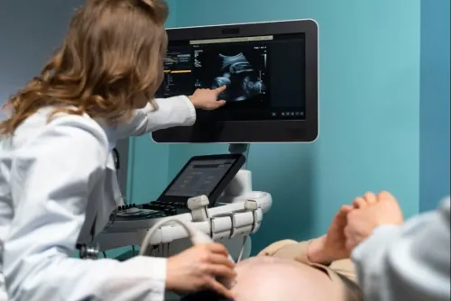
2. Know which approach is planned. A transvaginal scan uses a covered, lubricated probe in the vagina and is preferred for very early pregnancy, cervical-length measurement and suspected ectopic pregnancy because it sits closer to the uterus and cervix. A transabdominal scan uses the probe over your abdomen and becomes more common as pregnancy advances.
The operator may use both if one view does not answer the clinical question.
3. Allow extra time at an antenatal ultrasound clinic in Mumbai. Appointment length changes with gestational age, fetal position, maternal body habitus and the need for targeted views, so a repeat view or consultation may extend the visit.
4. Bring the referral or prescription, previous ultrasound reports, blood-test results, your last-menstrual-period date or IVF transfer details, and a symptom and medical-condition list. Before an obstetric ultrasound scan in Mumbai begins, mention multiple pregnancy, prior miscarriage, bleeding, pain, diabetes, hypertension, reduced movements, suspected placenta previa or breech presentation.
These details change image interpretation and the next step.
Choose a Mumbai clinic by capability, not by scan package
The answer to which obstetric ultrasound scan is right for me in Mumbai depends on gestational age, symptoms, pregnancy risk and the clinical question—not on a 3D or 4D label. Compare the antenatal ultrasound clinic in Mumbai by what it can assess and how it reports the result.
CheckWhat to verifyWhy it mattersOperatorAsk who performs and interprets the examinationSkill, equipment and clinical context affect accuracyProtocolConfirm it matches your gestational age and prescriptionA growth scan cannot replace an anomaly or targeted scanReportLook for fetal number, gestational age, measurements, placenta, amniotic fluid and the requested assessmentMissing details can make obstetric decisions harderGrowth studyRequire biparietal diameter, head circumference, abdominal circumference, femur length and estimated fetal weightOne estimated weight is not an exact weightDopplerAsk whether Doppler indices will be included when placental dysfunction or growth restriction is suspectedIt is not a routine add-on for every uncomplicated pregnancyFollow-upConfirm how findings are explained and whether the clinic coordinates with your obstetrician or refers to fetal medicineStructural or cardiac concerns need appropriate review
A legitimate Mumbai provider must comply with the Pre-Conception and Pre-Natal Diagnostic Techniques (Prohibition of Sex Selection) Act and associated rules, including registration, records, consent and required displays. Sex selection and communicating fetal sex for non-medical purposes are prohibited.
Dr. SPENTA's GYNAEC CLINIC can help when symptoms, IVF conception, twins or a previous report make scan selection less straightforward. Take your prescription and earlier records so the recommendation reflects your pregnancy, not an advertised package.
Related services
OBSTETRICS
An ultrasound is a procedure that uses sound waves to create images of your baby while it’s in the uterus.
View service →OBSTETRICS
can identify whether your baby is more or less likely to have any birth defects or genetic disorders like Down syndrome, Chromosonal abnormalities,...
Frequently asked questions
- Which obstetric ultrasound scan is needed first?
- A dating and viability scan is appropriate when your last menstrual period is unknown or unreliable, cycles are irregular, conception followed IVF, or you have pain or bleeding. Before about 10 weeks, a transvaginal scan often shows the uterus and embryo more clearly than an abdominal scan.
- What can different pregnancy screening tests detect?
- Screening tests answer different questions. Dating scans estimate gestational age and confirm location and viability; first-trimester screening assesses the risk of selected chromosomal conditions; and the anatomy scan examines fetal structures. A screening result estimates risk and does not diagnose every condition.
- When are anatomy, growth and Doppler scans appropriate?
- The anatomy scan assesses fetal structures at the recommended mid-pregnancy stage. Growth scans measure size and growth over time, while Doppler scans assess blood flow when a clinical reason exists, such as concern about placental function or fetal growth.
- How should I prepare for an obstetric ultrasound scan in Mumbai?
- Follow the clinic’s instructions about bladder filling, especially for early abdominal scans, and bring your last menstrual period date, IVF or conception details, previous reports, blood-group information and medication list. Tell the sonologist about pain, bleeding, prior pregnancy complications and relevant medical conditions.
Keywords